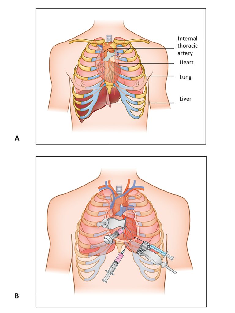
Pericardiocentesis
Pericardiocentesis is a medical technique that involves draining fluid from the pericardial sac, which is a double-walled sac that surrounds the heart. Normally, the pericardial sac contains a little quantity of fluid that functions as a lubricant, allowing the heart to move easily throughout the chest cavity. Excessive fluid buildup (pericardial effusion) can occur for a variety of reasons, including infection, inflammation, trauma, malignancy, or heart surgery.
Pericardiocentesis is used to alleviate pressure on the heart caused by extra fluid and to determine the origin of the effusion. Here’s a rundown of the phases:
Procedure:
Patient Positioning: To make the patient comfortable, the treatment is frequently conducted under local anesthetic with sedation.
Continuous monitoring of vital indicators such as heart rate, blood pressure, and oxygen saturation is required during the process.
Sterile Preparation: The skin above the pericardial sac is cleansed and sterilized, generally on the left side of the chest.
Local Anesthesia: To numb the region where the needle will be put, a local anesthetic is injected into the skin and underlying tissues.
Needle Insertion: Under imaging guidance, such as echocardiography or fluoroscopy, a needle is delicately introduced through the chest wall into the pericardial sac.
Excess fluid is removed from the pericardial sac after the needle is in the proper place. To ascertain the cause of the effusion, the fluid may be submitted for analysis.
Monitoring: During the surgery, the patient’s vital signs and symptoms are constantly monitored to detect any problems.
After draining the fluid, imaging may be conducted again to examine the quantity of fluid remaining and to confirm that the surgery was effective.
Complications:
While pericardiocentesis is usually regarded as a safe surgery, there are certain risks involved, including:
Infection from Bleeding
Damage to surrounding structures (for example, the heart, lungs, or blood arteries)
Indications:
Cardiac Tamponade: When there is fast fluid buildup causing compression of the heart (cardiac tamponade), leading to reduced cardiac function, pericardiocentesis is frequently done.
Pericardiocentesis is also performed to acquire a sample of pericardial fluid for diagnostic purposes, such as the identification of infections, cancer cells, or other abnormalities.
Lifestyle Considerations:
Individuals who have had pericardiocentesis may need to make certain lifestyle changes and follow particular protocols to enhance recovery, minimize problems, and limit the chance of recurrence.
Follow-up:
Following the surgery, patients may be evaluated for evidence of pericardial effusion recurrence or other problems. If not previously recognized, the underlying cause of the effusion will be examined and addressed.
In some medical settings, pericardiocentesis is a vital and potentially life-saving operation that is normally performed by qualified healthcare experts such as cardiologists or interventional radiologists.

